


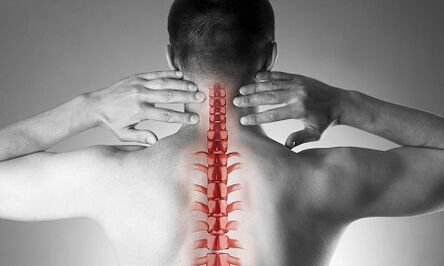
The cervical osteocondrosis, or spondylosis, occurs following changes in the shape and structure of the vertebrae.Despite the fact that the cervical region is quite short compared to the total length of the spine, it is perhaps the most important part of the spinal column.Each pair of close vertebrae forms the intervertebral holes through which the nerve roots go and go to each muscle and organ of the upper half of the body.Through other holes - in the side processes of these vertebrae - the vital vessels ensure the flow of blood to the brain.
The causes of osteochondosis of the cervical column
The causes of osteocondrosis are:
- injuries,
- "Sedentary" work to the monitor located below the eye level,
- physical work associated with the transfer of weights,
- In the long term he remains to guide a car,
- He works "on the phone" without the use of remote devices (in this case, the operator presses the phone on the ear shoulder)
- Constitutional characteristics (congenital changes of crooked in the cervical vertebrae, short neck)
Training of pathological vertebral changes
With osteochondrosis, small points begin on the edges of the vertebral bodies, which can hurt the structures located nearby.Very often, this occurs in response to an excess load on the cervical compartment, and not only the result of the "aging" of the intervertebral joints (remember that it was used to be considered the degenerative osteochondosis, therefore a natural disease "related to age", such as osteoarthritis).As the disease develops, the closing plates of the vertebrae occur and a decrease in the height of the intervertebral discs.These discs are normal play the role of the chip between the vertebrae and, among other things, prevent damage to the spinal roots.With progressive osteochondrosis, a protruding (hernia) of the nucleus of the intervertebral disc jacket occurs, on which during the disease there is more and more pressure while weakening "retaining" ligaments from all sides.This hernia is also able to squeeze spinal structures and cause neurological manifestations of the disease.
What are the symptoms of cervical osteochondosis?
Osteochondrosis of the cervical column with pain syndrome
Any pain in the neck forces the pathology of the cervical column.In terms of growth, the intensity of pain syndrome is divided into 4 phases, the first patient feels numb, tingling, a sense of "tension" in the area of a certain muscle group, in the fourth phase - the most serious - the pain is so intense that they lead to the immobility of the patient and the loss of performance.
In addition to pain syndrome in the cervical and occipital region, the patient notes the "reflected" pains (radiant) in the upper limb, the secondary areas of the chest chest.
Osteochondrosis of the cervical column with root syndrome
They speak of the involvement in the process of the nerve roots when pain, numbness and tingling spread to the lower jaw, the upper part of the back, the forearm and the fingers.At the same time, the patient attracts attention to the fact that "he seemed to leave" his hand, he slept uncomfortable.You can see the morning rigidity in the joints of the fingers, which do not last more than 10-15 minutes.With the development of root syndromes, during the examination, you can see a decrease in the muscle strength of the higher ends.
Osteochondrosis of the cervical column with the "vertebral artery syndrome"
Information on involvement in the process of blood vessels (tightening them with Ernian or osteophyte protruding), they say that when the patient complains of frequent attacks of headaches, especially after a long stay in a certain position, when he is thrown out of his head (for example, when he swims with a brass), if the noise in the ears and the dizzying are worried.This clinical situation is well detected using ultrasound (with the "Doppler mapping regime").With the ultrasound, the inquisition of the vertebral arteries, the narrowing of their light is determined.In this case, we can speak of surgery, since a pronounced change in the blood flow in the vertebral arteries is a risk factor for the development of the stroke.
Osteochondrosis of the cervical column with "cardiac syndrome (heart)"
This syndrome forces the patient to contact the cardiologist mainly, since the main complaints refer to the pain in the left half of the chest, to the sub -phapular region, which weaken or intensify when physical activity or body position is performed.After the exclusion of the infarction of the myocardium and other heart disease, the patient falls within the observation and treatment of a neurologist and orthopedist.
Diagnostics
To clarify the diagnosis, four methods are used: radiography, ultrasound, computerized tomography and magnetic resonance imaging.
The most convenient method is still the radiography of the cervical column, the most information is the radiography in the lateral projection ("lateral view").This method allows the first approximation to establish the presence of injuries, serious structural changes in the vertebrae.
The examination of ultrasound (ultrasound) is performed to clarify the condition of the vertebral arteries.With the help of this method, they discover whether the blood flow is disturbed and, in this case, to what extent and what type of obstacles have arisen and where they are located.
Computerized tomography (CT).It allows you to evaluate the state of bone structures more accurately, the degree of bone density, allows you to see the smaller osteophytes (bone exterior) than it is possible with X -raggi.
Magnetic resonance imaging (MRI).This type of exam is essential for suspicious hernias, an accurate location of the damage to the spinal cord and the degree of this damage.This study is necessary if the question is raised by the surgical (surgical) treatment of diseases of the cervical column.
Treatment of cervical osteochondosis
Pharmacological treatment
The standard series of products for the treatment of cervical osteochondosis reflects the purpose of the treatment: relieving pain syndrome, removing painful muscle spasm and inflammation of the nerve roots, increasing the mobility of the spine.To achieve these objectives, mainly the use of painkillers, fans -non -pound -shaped anti -anti -inflammatory flames, muscle relaxant are used.It should be remembered that the car -Media from these groups can be dangerous, since there is a possibility of incorrect interpretation of the symptoms, as well as underestimation of the side effects of these drugs.Local drugs (Basel) among fans in the form of gel are widely used and if the pain is stopped, the same drugs can already be used in the form of ointments.
For the treatment of osteocondrosis at a deeper, basic level ", systemic drugs are used. These substances restore the cartilage structures of the vertebrae, prevent their further damage. The treatment courses are long, the effect persists for many months.
Cervical osteochondrosis has significant differences compared to the pathology of the other spine.The pain in the neck in this case cannot be caused by the signals of the spinal nerves suffering, but by painful strange of chronic muscle - all together it is called muscle tonic syndrome.This is a completely "benign" state, which is well cared for with the same series of drugs: non -pounded anti -anti -inflammatory drugs, muscle relaxants, using an intramuscular "block" using steroids.Usually, the doctor reveals severe pain when the points called "trigger" along the entire cervical column, as well as in the muscles of the upper shoulder strap muscles.More often such a pathology occurs in women, mostly under the age of 40.Despite the pronounced pain syndrome, the vascular-north structures remain intact, the blood flow of the head area does not suffer.
Manual therapy
This method of treatment can be effective for the recently deriving pain (often due to a small injury, subluxation), not accompanied by dizziness, other changes from the nervous system and the circulatory system.It is allowed to resort to manual therapy only after an in -depth examination, moreover, the doctor who performs this procedure should have sufficient experience in the field of traumatology and orthopedics.With "old" shapes of the disease, the use of manual therapy is dangerous!
Two methods of this type of intervention are known:
- manipulation (clear short influences of a significant force aimed at eliminating subluxation, well known "bone clicks");
- Mobilization (the method is based on a stroke of the smooth neck after heating and relaxing the muscle corset of the neck).
A combined method is also used based on a combination of two main ones.It is important to remember that in addition to these contraindications, manual therapy is prohibited for any disease, accompanied by an increase in blood pressure, for any pathology of the thyroid gland and the anet or the organ.
Treatment of cervical osteochondosis at home
Medical gymnastics for cervical osteochondosis
The first and main rule for beginners to engage in physiotherapy exercises is not to perform exercises, overcoming painful sensations.Of course, you shouldn't start in the "acute" period in which pain has just appeared.Another important recommendation is to avoid sudden movements and circular movements in the cervical region.
Each lesson must start with a car -smashing the short light of the neck muscles.
What follows is a hot "heating" -Up:
- The hands are lowered along the body, the shoulders are uniform, the rear is straight (you can control the posture slightly pressed with heels, shoulder blades and buttocks on the wall).We walk instead of 1 minute on the whole foot, 1 minute - on socks, 1 minute - on the heels.
- The starting position is the same.We tighten the brushes in the punches, that they like their backs, our hands are straightened.The movements are slow, we make 20 repetitions, the last increase is more than 5 seconds.We make sure that the neck muscles are not "blocked".
- The starting position is the same.We tilt your head in turn to the right, then on the left side.The movements are smooth, one slope out of 8 accounts, at the extreme point of the inclination - hold for 8 seconds.
- The starting position is the same or sitting in a hard chair.Smooth inclinations of the head forward, at the extreme point - hold for 8 seconds
- The starting position is the same or sitting in a hard chair.Slowly incur your head forward, until the chin in the chest, then slowly turn your head to the right (at 4 accounts) and left (at 4 accounts).Do not allow muscle tension.
- The starting position is the same or sitting in a hard chair.We lift our backs on 4 accounts, so we also lower them in 4 counts.10 repetitions.
- The starting position is the same or sitting in a hard chair.We breed our backs, but now we perform circular movements in front of the back, 8 accounts.10 repetitions.
- We align the back, check the posture.At 4 accounts, we reduce the shoulder blades behind the back, trying to connect them, at the end that we roll up for 8 seconds, then we return to the starting position.
Cushions
As already mentioned, the hypertonicity of the muscles of the neck is the first and often the main reason for the development of cervical osteochondosis.A rational selection of cushions and mattresses, guaranteeing a relaxed and comfortable position during sleep is no less than gymnastics, accbourse and drugs.
When choosing a mattress, pay attention to the composition of the filling (the products are suitable, at least half made of coconut chips, that is, with a sufficient degree of rigidity).The soft spring mattresses do not provide sufficiently by straightening the spine.Most optimal sleep for sleep is on the side, pull one or both knees in the stomach.The pillow should be found in such a way as to fill the entire space between the shoulder, the ear and the mattrati, the parietal part (crown) of the head is on the same horizontal line with the spine.To avoid too high and too low, as well as soft cushions.The ideal option is a product of an ergonomic form, that is, in this case, with a small narrow on one side.
General recommendations
Pay attention to posture.During the walk or in the erect position, the location is a position when the chest protrudes forward and the stomach is pulled.
Avoid long -term in a sitting position.A simple rule of prevention of cervical osteochdrosis is known: after every 60 minutes of work, a period of 10-15 minutes of walking or heating is required.
A job chair should have a headrest or back.
In a sitting position, the legs should rest on the floor and the neck should not be tense.For this purpose, use special orthopedic devices: rollers under the neck while driving in a car, a pillow under the rear.
Avoid lifting weight.If necessary, kneel, press the heavy object on the body and then stopped using the strength of the leg muscles, but not the "push" of the back.
Don't lean with straightened legs.Use the stands, the work surfaces to bring the object closer to yourself and not to convince your face to the subject.Try to do the homework sitting on a chair or a gym ball.
If you need to use a mop, a broom or rake, do not stray your arms, back and neck, do not lean on laterally.
Avoid swimming in the brass style.